New research unveils morphological predictors in brain metastases
Tuesday August 20, 2024
Stereotactic radiotherapy is the one of the preferred treatments for patients with fewer than five brain metastases (BMs). However, some lesions recur after irradiation. A MOLAB study in collaboration with Fundación Instituto Valenciano de Oncología, Hospital de San Chinarro, MD Anderson Cancer Center, Hospital Regional Universitario de Málaga and Salamanca University Hospital, at Spain, intended to identify patients who are at a higher risk of failure, which can help in adjusting treatments and preventing recurrence.
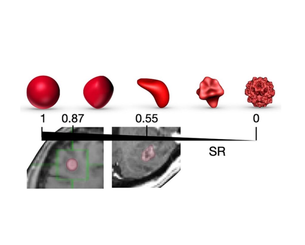
In the retrospective multicenter study, the researchers analyzed the predictive significance of a set of interpretable morphological features derived from contrast-enhanced (CE) T1-weighted MR images as imaging biomarkers using Kaplan-Meier analysis. The feature sets studied included the total and necrotic volumes, the surface regularity and the CE rim width. Additionally, they evaluated other non-morphological variables with potential prognostic value.
A total of 183 lesions in 128 patients were included in the study. Interestingly none of the studied variables measured at diagnosis were found to have prognostic value. However, the total and necrotic volumes and the CE rim width measured at the first follow-up after treatment and the change in volume due to irradiation can be used as imaging biomarkers for recurrence. The optimal classification was achieved by combining the changes in tumor volume before and after treatment with the presence or absence of necrosis (p < < 0.001).
Thus, the study demonstrated the prognostic significance of interpretable morphological features extracted from routine clinical MR images following irradiation in brain metastases, offering valuable insights for personalized treatment strategies.