

Metabolic tumor burden assessed by dual time point 18F-FDG PET/CT in locally advanced breast cancer: Relation with tumor biology.
A.M. García-Vicente, J. Pérez-Beteta, A. Martínez-González, V.M. Pérez-García, D. Molina, G.A. Jiménez Londoño, A. Soriano-Castrejón
Molecular Imaging and Biology 19(4), 636-644 (2017)
MOLAB authors
Abstract
Aim:
To investigate the influence of dual time point 18F-FDG PET/CT in the SUV and volume-based metabolic variables of breast lesions and their relation with biological characteristics and molecular phenotypes.
Material and methods:
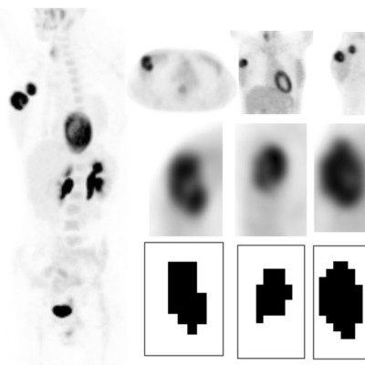
Prospective study including 67 patients with locally advanced breast cancer (LABC). All patients underwent a standard 18F-FDG PET/CT (PET-1) followed by a delayed acquisition (PET-2).
Tumors were segmentation following a three-dimensional methodology. Semi-quantitative metabolic variables (SUVmax, SUVmean and SUVpeak) and volume-based variables (metabolic tumor volume, MTV and total lesion glycolysis, TLG) were obtained.
Biologic prognostic parameters, such as the hormone receptors status, p53 and HER2 expression, proliferation rate (Ki-67) and grading were obtained. Molecular phenotypes and risk-classification [low: luminal A, intermediate: luminal B HER2 (-) or luminal B HER2(+) and high: HER2pure or triple negative] were established.
Spearman test was used to study the relation between clinical and biological variables with the metabolic parameters. The relevance of each metabolic variable in the prediction of phenotype risk was assessed using a multivariate analysis.
Results:
Most metabolic variables obtained in the PET-1 and PET-2 showed high and significant correlations between them. MTV and SUV variables (SUV max, SUV mean and SUV peak) where only marginally correlated.
SUV variables showed associations with hormone receptors status (estrogen, p<0.001 in all cases; progesterone, p=0.001 in all cases) and risk-classification according to phenotype (SUVmax, p=0.003; SUV mean, p=0.004; SUV peak, p=0.003). As to volume-based variables, only TLG showed association with hormone receptors status (estrogen, p<0.001; progesterone, p=0.031), risk-classification (p=0.007) and grade (p=0.036). Hormone receptors negative tumors, high grade tumors and high-risk phenotypes showed higher TLG values.
No association was found between the metabolic variables and Ki-67, HER2 or p53 expression.
Conclusion:
PET-1 SUV and volume-based variables were predictors of those obtained in PET-2. Most PET-derived parameters showed high association with molecular factors of breast cancer.